About the course
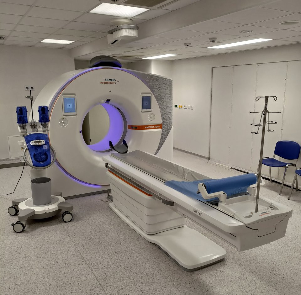
Build an introductory specialist understanding of computed tomography within an existing radiography practice route. The proposed certificate focuses on image formation, cross-sectional anatomy, patient care and quality awareness. Southern African cases should consider service capacity and communication, while keeping every patient-facing activity within an approved learner scope.
What you'll learn
- Explain the principles of CT image formation and common image limitations.
- Identify relevant cross-sectional anatomy in approved teaching images.
- Describe patient-protection and quality responsibilities.
- Document a supervised CT learning case with clear evidence and reflection.
Requirements
- This proposed specialist study assumes an appropriate existing diagnostic-radiography qualification, current professional registration and access to approved CT supervision. BMIT must confirm the actual entry route and scope with the relevant regulator. It is not presented as an initial route into radiographer registration.
Course content
Subjects in this programme
The subjects are grouped by learning stage, from foundations to specialist study and supervised application. Complete earlier foundations before the related advanced work. Clinical, laboratory, field and workplace subjects use approved facilities and supervision appropriate to the programme.
12 subjects are listed below, with a short description of each. The Subject descriptions tab contains the learning outcomes, topics, practical tasks and assessments.
Stage 1: Foundations
- Human Anatomy. Examine body organisation, musculoskeletal structures and their relationship within human anatomy. The subject develops thoracic structures and abdominal structures, then examines nervous structures and anatomical relationships. Study approved anatomical models and images in a supervised setting.
- Human Physiology. Develop your understanding of human physiology through homeostasis and cardiovascular function. The subject develops respiration and renal function, then examines endocrine regulation and nervous function. Interpret physiological teaching data and supervised non-invasive demonstrations.
- Diagnostic Imaging Physics. Study radiation interactions and image formation as foundations for diagnostic imaging physics. The subject develops equipment principles and digital imaging, then examines image quality and protection. Interpret approved teaching images and supervised equipment demonstrations.
- Health Ethics and Professional Practice. Examine consent, confidentiality and their relationship within health ethics and professional practice. The subject develops scope of practice and equity, then examines professional accountability and ethical reasoning. Discuss a simulated ethical dilemma with attention to patient rights.
- Clinical Communication. Develop your understanding of clinical communication through listening and plain language. The subject develops cultural context and shared information, then examines team handover and documentation. Practise communication in supervised simulation using fictional cases.
Stage 2: Technical and applied subjects
- CT Physics and Image Formation. Study acquisition principles and detectors as foundations for ct physics and image formation. The subject develops reconstruction and image parameters, then examines artefacts and quality evaluation. Analyse de-identified images and approved simulator data.
- CT Cross-Sectional Anatomy. Examine orientation, brain and their relationship within ct cross-sectional anatomy. The subject develops thorax and abdomen, then examines pelvis and musculoskeletal landmarks. Annotate approved CT teaching cases under qualified supervision.
- CT Clinical Practice. Develop your understanding of ct clinical practice through patient assessment context and examination planning. The subject develops contrast responsibilities and clinical workflow, then examines image review and handover. Complete approved CT observation and supervised practice within existing radiography scope.
- Radiation Protection. Study justification and optimisation as foundations for radiation protection. The subject develops exposure concepts and staff protection, then examines quality responsibilities and incident reporting. Analyse an authorised radiation-protection teaching case.
- Imaging Informatics. Examine digital images, image archives and their relationship within imaging informatics. The subject develops data standards and display, then examines privacy and workflow reliability. Use de-identified teaching data to review an imaging information workflow.
Stage 3: Supervised practice and integration
- CT Quality Assurance. Develop your understanding of ct quality assurance through quality indicators and equipment checks. The subject develops dose information and artefact review, then examines audit and improvement. Analyse approved CT quality data with the responsible imaging team.
- Integrated Supervised Clinical Practice. Study approved clinical roles and care coordination as foundations for integrated supervised clinical practice. The subject develops case reasoning and observed competence, then examines professional feedback and reflection. Complete an approved placement with qualified clinical supervision and authenticated records.
Assessment and practical learning
- Proposed assessment: written case analyses, knowledge assessments and discussion of evidence and professional responsibilities.
- Observed simulation followed by assessed practice at an approved clinical site, with an authenticated competence portfolio.
- An applied service or evidence-review project. Clinical and final assessment requirements must be approved by the education provider and relevant regulator.
Practical application
CT practical learning needs an approved imaging service, registered radiography supervisors and observed competence. Learners must not operate independently, alter clinical protocols or make diagnostic reports outside their authorised role. Online theory and image exercises do not replace clinical training or establish specialist registration.
Subject descriptions
Each subject below includes its purpose, learning outcomes, main topics, practical task and assessment approach.
Human Anatomy
Stage 1: Foundations
Examine body organisation, musculoskeletal structures and their relationship within human anatomy. The subject develops thoracic structures and abdominal structures, then examines nervous structures and anatomical relationships. Study approved anatomical models and images in a supervised setting.
Learning outcomes
- Explain body organisation and musculoskeletal structures using an appropriate example.
- Analyse a subject-related problem involving thoracic structures and abdominal structures.
- Present reasoned evidence addressing nervous structures and anatomical relationships.
Main topics
- Body organisation
- Musculoskeletal structures
- Thoracic structures
- Abdominal structures
- Nervous structures
- Anatomical relationships
Practical task
Study approved anatomical models and images in a supervised setting.
Assessment
Submit an annotated anatomy portfolio and identification assessment.
Human Physiology
Stage 1: Foundations
Develop your understanding of human physiology through homeostasis and cardiovascular function. The subject develops respiration and renal function, then examines endocrine regulation and nervous function. Interpret physiological teaching data and supervised non-invasive demonstrations.
Learning outcomes
- Explain homeostasis and cardiovascular function using an appropriate example.
- Analyse a subject-related problem involving respiration and renal function.
- Present reasoned evidence addressing endocrine regulation and nervous function.
Main topics
- Homeostasis
- Cardiovascular function
- Respiration
- Renal function
- Endocrine regulation
- Nervous function
Practical task
Interpret physiological teaching data and supervised non-invasive demonstrations.
Assessment
Submit a function-based case analysis and data interpretation.
Diagnostic Imaging Physics
Stage 1: Foundations
Study radiation interactions and image formation as foundations for diagnostic imaging physics. The subject develops equipment principles and digital imaging, then examines image quality and protection. Interpret approved teaching images and supervised equipment demonstrations.
Learning outcomes
- Explain radiation interactions and image formation using an appropriate example.
- Analyse a subject-related problem involving equipment principles and digital imaging.
- Present reasoned evidence addressing image quality and protection.
Main topics
- Radiation interactions
- Image formation
- Equipment principles
- Digital imaging
- Image quality
- Protection
Practical task
Interpret approved teaching images and supervised equipment demonstrations.
Assessment
Submit an image-physics and quality-analysis portfolio.
Health Ethics and Professional Practice
Stage 1: Foundations
Examine consent, confidentiality and their relationship within health ethics and professional practice. The subject develops scope of practice and equity, then examines professional accountability and ethical reasoning. Discuss a simulated ethical dilemma with attention to patient rights.
Learning outcomes
- Explain consent and confidentiality using an appropriate example.
- Analyse a subject-related problem involving scope of practice and equity.
- Present reasoned evidence addressing professional accountability and ethical reasoning.
Main topics
- Consent
- Confidentiality
- Scope of practice
- Equity
- Professional accountability
- Ethical reasoning
Practical task
Discuss a simulated ethical dilemma with attention to patient rights.
Assessment
Submit a justified ethical decision and professional reflection.
Clinical Communication
Stage 1: Foundations
Develop your understanding of clinical communication through listening and plain language. The subject develops cultural context and shared information, then examines team handover and documentation. Practise communication in supervised simulation using fictional cases.
Learning outcomes
- Explain listening and plain language using an appropriate example.
- Analyse a subject-related problem involving cultural context and shared information.
- Present reasoned evidence addressing team handover and documentation.
Main topics
- Listening
- Plain language
- Cultural context
- Shared information
- Team handover
- Documentation
Practical task
Practise communication in supervised simulation using fictional cases.
Assessment
Complete an observed communication exercise and reflective record.
CT Physics and Image Formation
Stage 2: Technical and applied subjects
Study acquisition principles and detectors as foundations for ct physics and image formation. The subject develops reconstruction and image parameters, then examines artefacts and quality evaluation. Analyse de-identified images and approved simulator data.
Learning outcomes
- Explain acquisition principles and detectors using an appropriate example.
- Analyse a subject-related problem involving reconstruction and image parameters.
- Present reasoned evidence addressing artefacts and quality evaluation.
Main topics
- Acquisition principles
- Detectors
- Reconstruction
- Image parameters
- Artefacts
- Quality evaluation
Practical task
Analyse de-identified images and approved simulator data.
Assessment
Submit a CT physics and image-quality report.
CT Cross-Sectional Anatomy
Stage 2: Technical and applied subjects
Examine orientation, brain and their relationship within ct cross-sectional anatomy. The subject develops thorax and abdomen, then examines pelvis and musculoskeletal landmarks. Annotate approved CT teaching cases under qualified supervision.
Learning outcomes
- Explain orientation and brain using an appropriate example.
- Analyse a subject-related problem involving thorax and abdomen.
- Present reasoned evidence addressing pelvis and musculoskeletal landmarks.
Main topics
- Orientation
- Brain
- Thorax
- Abdomen
- Pelvis
- Musculoskeletal landmarks
Practical task
Annotate approved CT teaching cases under qualified supervision.
Assessment
Complete an image-based anatomy assessment.
CT Clinical Practice
Stage 2: Technical and applied subjects
Develop your understanding of ct clinical practice through patient assessment context and examination planning. The subject develops contrast responsibilities and clinical workflow, then examines image review and handover. Complete approved CT observation and supervised practice within existing radiography scope.
Learning outcomes
- Explain patient assessment context and examination planning using an appropriate example.
- Analyse a subject-related problem involving contrast responsibilities and clinical workflow.
- Present reasoned evidence addressing image review and handover.
Main topics
- Patient assessment context
- Examination planning
- Contrast responsibilities
- Clinical workflow
- Image review
- Handover
Practical task
Complete approved CT observation and supervised practice within existing radiography scope.
Assessment
Submit an authenticated CT competence portfolio and case discussion.
Radiation Protection
Stage 2: Technical and applied subjects
Study justification and optimisation as foundations for radiation protection. The subject develops exposure concepts and staff protection, then examines quality responsibilities and incident reporting. Analyse an authorised radiation-protection teaching case.
Learning outcomes
- Explain justification and optimisation using an appropriate example.
- Analyse a subject-related problem involving exposure concepts and staff protection.
- Present reasoned evidence addressing quality responsibilities and incident reporting.
Main topics
- Justification
- Optimisation
- Exposure concepts
- Staff protection
- Quality responsibilities
- Incident reporting
Practical task
Analyse an authorised radiation-protection teaching case.
Assessment
Submit a protection review within the approved professional role.
Imaging Informatics
Stage 2: Technical and applied subjects
Examine digital images, image archives and their relationship within imaging informatics. The subject develops data standards and display, then examines privacy and workflow reliability. Use de-identified teaching data to review an imaging information workflow.
Learning outcomes
- Explain digital images and image archives using an appropriate example.
- Analyse a subject-related problem involving data standards and display.
- Present reasoned evidence addressing privacy and workflow reliability.
Main topics
- Digital images
- Image archives
- Data standards
- Display
- Privacy
- Workflow reliability
Practical task
Use de-identified teaching data to review an imaging information workflow.
Assessment
Submit a data-governance and workflow report.
CT Quality Assurance
Stage 3: Supervised practice and integration
Develop your understanding of ct quality assurance through quality indicators and equipment checks. The subject develops dose information and artefact review, then examines audit and improvement. Analyse approved CT quality data with the responsible imaging team.
Learning outcomes
- Explain quality indicators and equipment checks using an appropriate example.
- Analyse a subject-related problem involving dose information and artefact review.
- Present reasoned evidence addressing audit and improvement.
Main topics
- Quality indicators
- Equipment checks
- Dose information
- Artefact review
- Audit
- Improvement
Practical task
Analyse approved CT quality data with the responsible imaging team.
Assessment
Submit a quality-assurance investigation and justified improvement plan.
Integrated Supervised Clinical Practice
Stage 3: Supervised practice and integration
Study approved clinical roles and care coordination as foundations for integrated supervised clinical practice. The subject develops case reasoning and observed competence, then examines professional feedback and reflection. Complete an approved placement with qualified clinical supervision and authenticated records.
Learning outcomes
- Explain approved clinical roles and care coordination using an appropriate example.
- Analyse a subject-related problem involving case reasoning and observed competence.
- Present reasoned evidence addressing professional feedback and reflection.
Main topics
- Approved clinical roles
- Care coordination
- Case reasoning
- Observed competence
- Professional feedback
- Reflection
Practical task
Complete an approved placement with qualified clinical supervision and authenticated records.
Assessment
Pass direct clinical assessments and submit a reflective competence portfolio.
Instructors
Enrolment options
National Certificate in Computed Tomography
Build an introductory specialist understanding of computed tomography within an existing radiography practice route. The proposed certificate focuses on image formation, cross-sectional anatomy, patient care and quality awareness. Southern African cases should consider service capacity and communication, while keeping every patient-facing activity within an approved learner scope.
- Teacher: ImfundoSpace Administrator
- Enrolled students: There are no students enrolled in this course.


